Fluorophotometry
Fluorophotometry is the established method for quantitating the permeability of the blood-retinal barriers and the blood-aqueous barrier. By measuring the concentration profile of the tracer fluorescein within the ocular cavity, the dynamics of intraocular diffusion and elimination can be accurately monitored. The resulting determinations provide indications of the physiological and pathological state of the retinal vasculature, the pigmented epithelium, the choroid and the ciliary processes.
The objective and quantitative capabilities of fluorophotometry permit the detection of physiological changes very early in the course of certain diseases, and the monitoring of progress in treatment.
Fluorophotometry is a successfully employed research tool in both laboratory and clinical settings.
The Fluorotron Master and Fluorophotometry
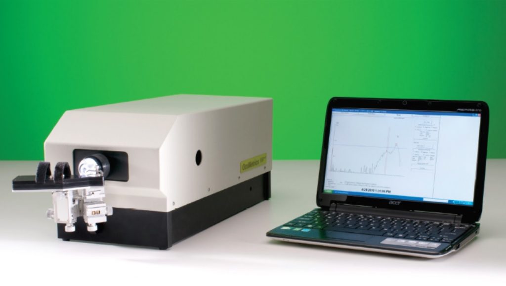
The OcuMetrics Fluorotron Master Fluorophotometer is a computerized system for measuring fluorescein within the eye and for processing the data into an easily accessible format. The Fluorotron constructs a fluorescein concentration profile by sequentially focusing on spatially distinct sites along the optical axis (from retina to cornea) of the ocular cavity. The data is processed by computer and displayed graphically. A hard copy can be obtained of both the graphic and the numerical data. By using the appropriate protocols, the posterior and anterior sources of leakage can be differentiated. This means that the blood retinal and blood-aqueous barriers can be independently monitored. The superior optical system offered by the Fluorotron produces accurate, reproducible results, making it the instrument of choice among foremost researchers in fluorophotometry.
The Fluorotron System
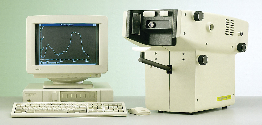
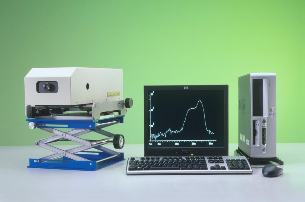
The Fluorotron system is made up of four major components; the electro-optic system (optic head), computer, display monitor, and printer.
The optic head is a fluorophotometer which delivers a specifically focused excitation beam of blue light into the ocular cavity and then receives the resulting fluorescent green light and directs it into a photodetector. By changing the focal plane every 0.25 mm, as many as 149 sequential readings are made along an axis from a position posterior to the retina to a position anterior to the cornea.
The operation of the optic head is controlled by the computer, prompting the operator through the entire procedure. The operator merely aligns the eye and initiates the scan. The scan is then automatically performed and displayed on the screen and printed in graphic and digital forms. Scan and patient data are stored on a magnetic fixed disk. The computer is used to process the data by using the appropriate program.
Versatile—Designed for vitreous fluorophotometry, endothelial permeability, aqueous flow studies, and other anterior segment studies.
Convenient—Provides computer storage of patient data and diffusion profiles for post-examination and future data processing, and for comparing changes over time.
Upgradable—Advances in fluorophotometry technology are readily incorporated into the system by changing disk software.
Accurate—Data processing by the computer corrects for in vivo artifacts.
Simple—All instrumentation operations are easily performed by a technician.
Self–calibrating—Automatic, internal calibration system.
FLUOROPHOTOMETER SPECIFICATIONS
| Optic Head | 2 mm at 3% peak signal |
| Depth of Resolution | 0.1 ng / ml fluorescein (3 x background fluctuations) |
| Sensitivity | 5% with solutions < 5 ng / ml |
| Reproducibility | 3% with solutions > 5 ng / ml |
| Laptop Computer | |
| OS | Windows 10 |
| Fluorotron Accessories: | The Fluorotron is supplied with a cuvette holder and two cuvettes which adapt to the optical head for measuring the concentration of fluorescein in blood plasma samples. |
| Options: | Anterior Chamber Adapter magnifies the scan for detailed work in the anterior chamber and lens. Anterior Chamber Diagnostic Protocol Software—a computer protocol for the manipulation of data specific to the anterior chamber and the computation of aqueous flow. |
NOT FOR HUMAN USE
Specifications subjects to change without notice.
Copyright 2021 OcuMetrics, Inc.
2224-C Old Middlefield Way
Mountain View, CA 94043-2421
(650) 960-3955
Fax: (650) 960-0611