The Standard in Ocular Research
Now Available for Use in Mice
Flurophotometry is the established method for quantitating the permeability of the bloodretinal barrier and the blood-aqueous barrier. By measuring the concentration profile of the tracer fluorescein within the ocular cavity, the dynamics of intraocular diffusion and elimination can be accurately monitored. The resulting determinations provide indications of the physiological and pathological state of the eye.
Fluorophotometry is a successfully employed research tool in both laboratory and clinical settings.
The Fluorotron Master and Fluorophotometry
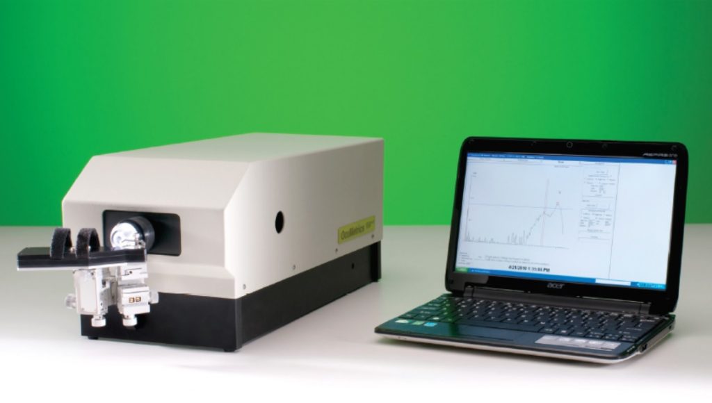
The OcuMetrics Fluorotron Master Fluorophotometer is a computerized system for measuring fluorescein within the eye and for processing the data into an easily accessible format. The Fluorotron constructs a fluorescein concentration profile by sequentially focusing on spatially distinct sites along the optical axis (from retina to cornea) of the ocular
cavity. The data is processed by computer and displayed graphically. A hard copy can be obtained of both the graphic and the numerical data. By using appropriate protocols, permeability and fluid turnover
in the eye can be determined. The superior optical system offered by the Fluorotron produces accurate, reproducible results, making it the instrument of choice among foremost researchers in fluorophotometry.
The Fluorotron System
The Fluorotron system is made up of four major components; the electro-optic system (optical head), computer, display monitor and
printer.
The optical head is a fluorophotometer which delivers a specifically focused excitation beam of blue light into the ocular cavity and then
receives the resulting fluorescent green light and directs it into a photodetector. By changing the focal plane a high resolution
concentration profile is measured along an axis through the eye.
The operation of the optic head is controlled by the computer, prompting the operator through the entire procedure. The operator merely aligns the eye and initiates the scan. The scan is then automatically performed and displayed on the screen and printed in graphic and digital forms. Scans and all data are stored on the computer hard drive. The computer is used to process the data by using the appropriate program.
Versatile—Designed for endothelial permeability, aqueous flow studies, and other anterior chamber studies.
Convenient—Provides computer storage of data and diffusion profiles for post-examination and future data processing, and for comparing
changes over time.
Upgradable—Advances in fluorophotometry technology are readily incorporated into the system by changing computer software.
Accurate—Computer data processing corrects for in vivo artifacts.
Simple—All instrumentation operations are easily performed by a technician.
Self-calibrating—Automatic, internal calibration system.
Scheduler
The Scheduler acts as an assistant to the operator. After a patient and protocol have been selected and a schedule has been created, the Scheduler keeps track of the time of day and will remind the operator to perform the next event when the time for that event arrives. This includes not only scans but also fluorescein instillation, rinsing, and other protocol related events. It can even be set to automatically initiate the scans.
“Smart Scan”
Many Fluorotron protocols do not need a full scan of the eye in order to calculate the correct results. These protocols use “Smart Scanning” to locate the area of interest and to measure only what is most relevant. This can speed up the time required for data acquisition considerably.
Enough context is measured around the area of interest so that the operator can be assured that the proper features are measured and that
the measurements are valid. About a dozen data points are measured for the Epithelial Permeability Protocol versus 149 for a full scan.
“Instant” Analysis
Landmark recognition is built into the program, so the analysis process is automatic. If the operator decides to edit out a data point or to change an experimental parameter, they merely have to press the on-screen “Update Analysis” button, and the results are recalculated and displayed in a fraction of a second.
References
Toris CB, Fan S, Johnson TV, Camras LJ, Hays CL, Liu H, Ishimoto BM. Aqueous Flow Measured by Fluorophotometry in the Mouse. Invest Ophthalmol Vis Sci. 57(8):3844-52 2016. PubMed Full Article
Colé N, Thoele J, Ullmer C, Foxton RH. Real-time measurements of vascular permeability in the mouse eye using vitreous fluorophotometry. Sci Rep 13(1):9226 (2023). PMC PDF
2224-C Old Middlefield Way
Mountain View, CA 94043-2421
(650) 960-3955
Fax: (650) 960-0611
sales@ocumetrics.com
www.ocumetrics.com
Not for use in humans.
Specifications subject to change without notice.
© 2020 OcuMetrics, Inc.